
Shape Completion in the Dark: Completing Vertebrae Morphology from 3D Ultrasound
Miruna-Alexandra Gafencu, Yordanka Velikova, Mahdi Saleh, Thomas Wendler, Mohammad Farid Azampour, Tamas Ungi, Nassir Navab
International Journal of Computer Assisted Radiology and Surgery (IJCARS), 2024
Abstract
Ultrasound imaging is attractive for spine assessment because it is radiation free, but clinicians only perceive a small visible window of the anatomy and must mentally recreate the occluded vertebrae. We introduce a point-cloud based probabilistic deep learning model that completes occluded lumbar structures from 3D ultrasound input. To train the network we mimic ultrasound physics to synthesize partial vertebrae point clouds and to model artifacts that typically hide the spinous process and facet joints. The approach produces anatomically consistent completions on synthetic and patient studies, with mean/median Chamfer Distance of 2.02 / 0.03. Ablations confirm that the physics-driven data generation is crucial (11.8 mean CD without it), and the method preserves key landmarks such as the spinous process (4.73 CD) and facet joints (4.96 mm mean distance to ground truth).
Highlights
- Probabilistic point-cloud network hallucinates the missing vertebra geometry while keeping visible ultrasound regions untouched.
- Training data is synthesized with a differentiable ultrasound simulator that injects realistic occlusions and attenuation patterns.
- Completion quality is validated on phantom and patient data, preserving anatomical landmarks with <5 mm error.
Method overview
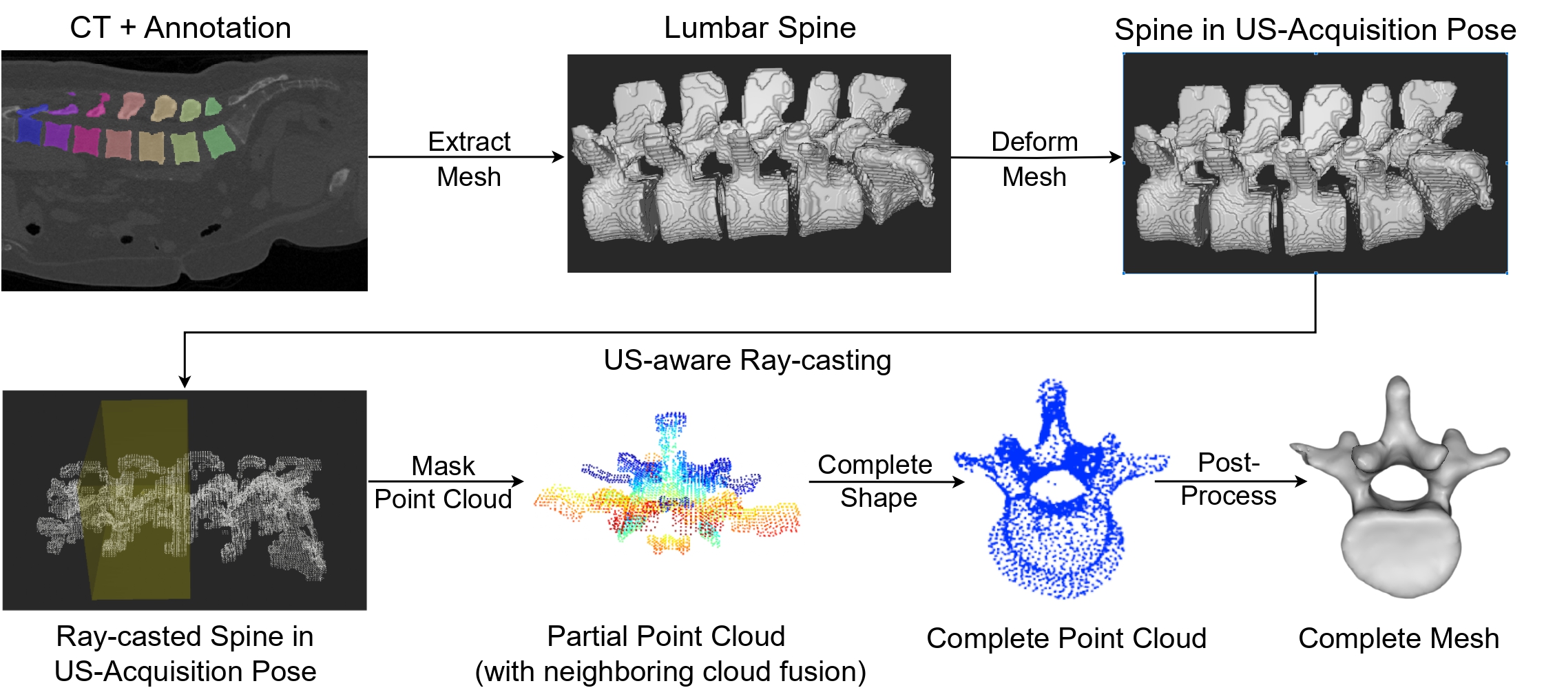
The framework decouples partial vertebra point extraction, probabilistic completion, and anatomical regularization. First, tracked 3D ultrasound sweeps are converted to point clouds annotated with their visibility map. These partial observations are fused with synthetic occlusions generated by simulating acoustic shadowing. The completion network predicts a distribution over the missing structure, enforces symmetry and level-aware features, and outputs a consolidated mesh with uncertainty estimates. This combination enables hallucinating hidden surfaces while maintaining patient-specific cues captured in ultrasound.
Results
Across synthetic vertebrae the model reaches a mean Chamfer Distance of 2.02 while maintaining a median error of 0.03 thanks to the probabilistic aggregation. On patient measurements the framework reconstructs spinous processes with 4.73 CD and facet joints with 4.96 mm distance, demonstrating that the completed models respect clinically relevant landmarks and injection sites even when the ultrasound signal is severely degraded.
Citation
If you reference this work, please cite:
@article{gafencu2024shapecompletion,
  title={Shape Completion in the Dark: Completing Vertebrae Morphology from 3D Ultrasound},
  author={Gafencu, Miruna-Alexandra and Velikova, Yordanka and Saleh, Mahdi and Wendler, Thomas and Azampour, Mohammad Farid and Ungi, Tamas and Navab, Nassir},
  journal={International Journal of Computer Assisted Radiology and Surgery},
  year={2024},
  doi={10.1007/s11548-024-03126-x}
}